- Special prices
- EEG
- EEG Caps
-
EMG | EP
- Cables and lines for EMG and EP devices
- EMG Needle Electrodes
- Subdermal needle electrodes for various applications
- Gold Cup Electrodes for heavy duty operations
- Cup Electrodes, EP, NLG and EMG
- Surface electrodes | Reusable | EMG and EP
- Surface electrodes | Adhesive electrodes for EMG and EP
- Stimulation Electrodes | EMG | EP
- EMG Bar Electrode bar / Block Electrode | Felt inserts
- Bar Electrode bar / Block Electrode | Gold-plated electrodes
- Accessories for Stimulation and Bar Recording Electrodes
- Ground Strap Electrodes
- Loop Electrodes
- BERA and AEP accessories and consumables
- Hearing test accessories | AEP and Audiometry
- Alligator Clips
- Nasal cannulas
-
Electrodes
- Natus | EEG and EMG accessories
- Nihon Kohden | Electrodes and Accessories
- EMG Needle Electrodes
- EMG Bar Recording Electrode
- Stimulation Electrodes
- Bridge Electrodes
- Cup electrodes and disposable cup electrodes
- Sensors | Sleep sensors | Spo2 sensors
- ECG Electrodes | ECG Adhesive Electrodes
- Subdermal Needle Electrodes
- Napf Electrodes of chlorinated sintered material
- Adhesive Electrodes for Neurological Examinations
- Surface Recording Electrodes / Waffle Electrodes
- Ground Electrodes
- Loop Electrodes
- Ear Electrodes
- IOM
- Cable
- Pastes | Gels
-
Sleep Lab
- Nasal Cannulas for screening and diagnostics in the sleep lab
- Alice 6 - accessories
- Cup Electrodes
- Gold Cup Electrodes
- Adhesive electrodes for sleep studies
- Snap Button Cables
- Oxygen Saturation Sensors
- Respiratory Sensors
- Airflow Pressure Sensors
- Body position sensors for detecting the sleeping position
- Respiratory Efforts
- Schnarchsensoren und Mikrofone
- More products
TerniMed Start Page Useful Information RIP- Respiratorische Induktive Plethysmographie
RIP- Respiratorische Induktive Plethysmographie

What is respiratory inductive plethysmography - RIP?
Respiratory inductive plethysmography (RIP) is a (non-invasive) method of monitoring respiration during sleep studies in the sleep laboratory.
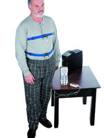
Elastic bands are placed around the thorax and abdomen, and an induction loop incorporated into these bands serves as a sensor that senses the respiratory movements of the thorax and abdomen.
These induction bands are very sensitive transducers for detecting volume changes in the parts of the body enclosed by the belts (thorax and abdomen).
In sleep medicine, this method is used to measure respiratory volume, respiratory rate, and monitor apnea, hypo- and hyperventilation.
With inductive plethysmography, it is possible to derive the relative volume changes within the respiratory system from the body surface movements necessary for breathing. Here, the elastic straps as a whole serve as a sensor (induction loop incorporated into the straps).
The induction belts placed around the thorax and abdomen register the circumferential changes of the two compartments, i.e. thorax and abdomen.
During inspiration, inhalation, the circumference increases due to the lifting of the thorax and the expansion of the abdominal wall, while during expiration, exhalation, the circumference decreases.
Measurement of circumference change is achieved by recording small changes in coil induction, in the RIP bands.
Technically, this is accomplished by varying the inductance of the (RIP) bands relative to the circumference of the body part they surround. The changes in inductance are then converted to an analog signal in the interface.
The sum signal (SUM) of the two individual compartments (thorax and abdomen) reflects the relative change in air volume in the airways.
This ensures that a correct signal is delivered regardless of the patient's body position, which may result in strain relief of part of the belt.
When using piezo-based sensors, as they are still used in many laboratories, a strain relief of the belt leads to a failure of the signal.
In respiratory inductive plethysmography, the sum signal of abdomen and thorax is approximately equal to the respiratory volume measured with a pneumotachograph.
© Ternimed
Contact UsWolfram Droh GmbH • Dammweg 11 • 55130 Mainz • Deutschland |
|||
Payment methodes
|
Socialmedia
|
||


